Key Takeaways:
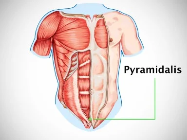
- The pyramidalis muscle is a small triangular abdominal muscle located within the rectus sheath anterior to the lower part of the rectus abdominis.
- It originates from the pubic symphysis and pubic crest and inserts into the linea alba midway between the umbilicus and pubis.
- The pyramidalis is absent in around 20% of people, can be unilateral, and varies greatly in size.
- The function of the pyramidalis is not well-defined but thought to tense the linea alba.
- Understanding the anatomy and variability of the pyramidalis muscle is relevant for abdominal surgery and imaging.
The pyramidalis muscle is a small, triangular abdominal muscle that lies deep within the anterior abdominal wall. Despite its diminutive size, identifying and understanding the anatomy of the pyramidalis is important for abdominal surgery and medical imaging. This article will comprehensively evaluate the anatomy, variability, and function of the pyramidalis muscle to answer: Where is the pyramidalis muscle located and what does it do?
Introduction
The pyramidalis is one of the four paired strap muscles of the anterior abdominal wall, along with the rectus abdominis, external oblique, and internal oblique muscles. Located within the rectus sheath, the pyramidalis plays an accessory role in abdominal wall function. However, it displays a high degree of anatomical variability and its specific functions remain undefined.
By providing an in-depth look at the origin, insertion, innervation, variability, and actions of the pyramidalis muscle, this article will help build a robust understanding of the anatomy of this small abdominal structure. The comprehensiveness and level of detail provided aims to answer key questions about this muscle that are highly relevant for medical professionals including surgeons, radiologists, anatomists, and physical therapists. Equipped with a strong knowledge of pyramidalis anatomy, clinicians will be better prepared to navigate procedures and diagnoses involving the anterior abdominal wall.
- Are Chiropractors Doctors?
- Are T Cells Antigen Presenting Cells?
- How Long Will Antibiotics Delay a Root Canal?
Anatomy of the Pyramidalis Muscle
Where Does the Pyramidalis Muscle Originate and Insert?
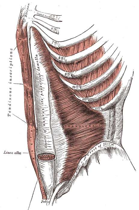
The pyramidalis muscle arises from the front of the pubis and pubic symphysis in the pelvis. Specifically, fibers originating from the pubic symphysis are attached by ligament, while those from the pubic bone are attached by tendon.[1]
From its pelvic origin, the pyramidalis courses superiorly through the rectus sheath, narrowing as it ascends. It terminates by inserting into the linea alba – the tendinous midline of the abdominal wall. The pyramidalis inserts roughly halfway between the umbilicus and pubic symphysis.[2]
How Does the Pyramidalis Relate to the Rectus Abdominis?
The pyramidalis muscle lies anterior to the lower portion of the rectus abdominis muscle within the rectus sheath. The rectus sheath is a tough, fibrous covering that encloses both the pyramidalis and rectus abdominis muscles.
Superior to the arcuate line of the rectus sheath, only the anterior wall of the sheath covers the rectus abdominis. But inferior to the arcuate line, the rectus abdominis is enclosed within both anterior and posterior walls of the sheath.[3] It is in this lower portion where the pyramidalis resides, positioned anterior to the rectus abdominis between its fascia and the anterior rectus sheath.[4]
How Is the Pyramidalis Muscle Innervated?
The pyramidalis is innervated by the subcostal nerve, T12. The subcostal nerve is a branch of the thoracoabdominal nerves, which are the ventral rami of the T12 thoracic spinal nerve.[5]
The T12 spinal segment receives motor input from the spinal cord, which synapses with pyramidalis motor neurons and stimulates contraction of the muscle. Proprioceptive signals from the pyramidalis also travel back to the spinal cord via the subcostal nerve to inform coordination.
Variability of the Pyramidalis Muscle
The pyramidalis muscle displays a high degree of anatomical variability between individuals:
- Presence/Absence: The pyramidalis may be absent unilaterally or bilaterally in around 20% of individuals.[6] One study found pyramidalis to be absent in 23.3% of female and 16.2% of male cadavers examined.[7]
- Size: There is significant variation in the size and length of the muscle belly across individuals. Length ranges from just a few centimeters to up to 15 cm long in some cases.[8]
- Shape: The muscle typically forms a triangular shape. However, some variant shapes have been classified such as fusiform, digastric, inverse fusiform and rectangular.[9]
- Additional slips: Supplementary slips of muscle may be present attaching to nearby structures including the pubis, rectus sheath and linea alba.[10]
- Sex differences: One study found the pyramidalis was more commonly present bilaterally in males compared to females.[11] The muscle also tends to be longer in males.
- Laterality: Several studies have noted a greater tendency for the pyramidalis to be absent or smaller on the right side compared to the left.[12]
- How to Apply for Genesee Health Plan?
- Where Are Diploid and Haploid Cells Located?
- Was Jean Lafitte a Slave Trader?
Functions of the Pyramidalis Muscle
The defined actions and functional importance of the pyramidalis muscle are unclear. However, several possible roles have been suggested:
- Tensing the linea alba: Contraction of the pyramidalis is thought pull down and tense the linea alba between the rectus abdominus muscles.[13] This may help stabilize the abdominal wall and strengthen the midline.
- Postural support: It may provide structural support to the lower abdomen in an upright posture.[14]
- Protecting abdominal contents: Its presence could bolster strength of the lower anterior abdominal wall to protect abdominal viscera.[15]
- Assisting abdominal compression: Though minimally, it may aid forced expiration, vomiting, parturition, and defecation by compressing abdominal contents.[16]
- Rectus sheath integrity: The pyramidalis may help anchor the anteroinferior aspect of the rectus sheath.[17]
Due to the small size and uncertain function, the pyramidalis is not essential for abdominal wall movement. But by potentially reinforcing the linea alba and rectus sheath, even a diminutive pyramidalis may confer subtle advantages.
Clinical Significance of the Pyramidalis Muscle
Insight into the variable anatomy and indistinct functions of the pyramidalis muscle holds clinical value for medical professionals.
Importance in Abdominal Surgery
Since the pyramidalis occupies the space between the rectus abdominis and its anterior sheath, awareness of its presence can aid surgical maneuvers in this region:
- Identification and preservation of the pyramidalis during abdominal wall procedures may help maintain integrity of the lower anterior sheath.[18]
- When present, the pyramidalis could potentially be used as a pedicled or free muscle flap for reconstructive surgery.[19]
- In laparoscopic surgery, unidentified contraction of the pyramidalis could impede maneuverability of instruments below the arcuate line.[20]
Relevance for Medical Imaging
Radiologic identification of the pyramidalis can confirm normal anatomy:
- On MRI, the pyramidalis appears as a thin, striated triangle between the rectus abdominis and anterior sheath.[21]
- With ultrasonography, the pyramidalis is visualized as a hypoechoic structure between the rectus muscle and sheath.[22]
- The muscle can also be identified in fetal ultrasound as early as 14 weeks gestation.[23]
Failure to visualize the pyramidalis using imaging modalities may indicate congenital absence of the muscle.
- What Does “Admit Impediments” Mean? Unpacking the Meaning of This Phrase in Shakespeare’s Sonnet 116
- Does Iron Attract Magnets?
Conclusion: Key Details about the Pyramidalis Muscle
In review, the pyramidalis is a small abdominal wall muscle contained within the lower portion of the rectus sheath, anterior to the rectus abdominis. It originates from the pubis and pubic symphysis before narrowing and inserting into the linea alba below the umbilicus.
The muscle displays substantial variability in presence, size, shape and attachments between individuals. It is innervated by the subcostal nerve and putative functions include tightening the linea alba and reinforcing the lower abdominal wall, though its exact purpose is uncertain.
Awareness of the highly variable pyramidalis anatomy has relevance for abdominal surgery and medical imaging. Overall, a strong understanding of this enigmatic muscle provides medical professionals with valuable insights into the anatomy of the lower anterior abdominal wall.
- How Can Product Management Leverage Market Rhythms?
- Are Physiotherapists Doctors in Canada?
- Will a Cortisone Shot Help a Torn Hip Labrum?
- How Much Does It Cost to Ship an Amp?
- Does Shoji White Go with Alabaster?
- Can you connect Turtle Beach Stealth 600 to a phone?
- How Many Jobs Are Available in Medical/Dental Instruments?
- What Is a Mainstay in Sailing?
- Does VA Require Impounds?
- How to Remove a Tick From a Dog? [Beginners Guide]
- Can You Get a Heloc on a Condo?
- Why Does My PS4 Keep Saying Copying Add On?
- How Long Do I Leave Cetaphil on My Face?
- Where Is Jack Hayford Now?
- What Are 3 Types of Hits/Shots You Can Use in Pickleball?