Key Takeaways:
- The retropharyngeal lymph nodes are located in the retropharyngeal space behind the upper pharynx.
- They lie between the buccopharyngeal fascia anteriorly and the prevertebral muscles posteriorly.
- The retropharyngeal space is found posterior to the pharynx and esophagus.
- The paired retropharyngeal lymph nodes are separated from the atlas vertebra by the longus capitis muscle.
- They are located medial to the internal carotid arteries and divide into medial and lateral masses.
- The lateral retropharyngeal lymph nodes sit just medial to the carotid sheath.
- Which Is Left Hand?
- How Much Is Cooltone?
- How Does a Weight Loss Club Effectively Improve Community Health?
Introduction
The lymphatic system plays a crucial role in immune function, fluid balance, and fat absorption. Lymph nodes are important structural components of this system located throughout the body. But where exactly are the retropharyngeal lymph nodes positioned? Understanding the anatomy of lymph nodes in different regions of the neck and head is key for medical professionals. This article will comprehensively evaluate the precise location and anatomical relations of the retropharyngeal lymph nodes.
Detailed knowledge of retropharyngeal lymph node location is essential for head and neck surgeons, radiologists, oncologists, and other specialists. It aids in accurate diagnosis of diseases involving these lymph nodes, technique and safety of surgical procedures, and optimal targeting for biopsy, excision, or radiation therapy. Furthermore, variations in lymph node location among individuals emphasize the need for in-depth understanding of the common patterns.
By the end of this article, readers will have a deep understanding of the retropharyngeal space boundaries, fascial layers, and nearby structures that define the anatomical position of the retropharyngeal lymph nodes. The level of detail provided aims to elucidate this anatomy and equip medical experts with knowledge for clinical practice.
What Is the Retropharyngeal Space?
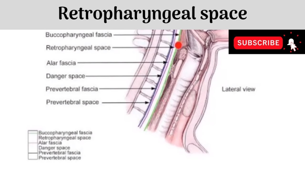
To comprehend retropharyngeal lymph node location, we must first understand the retropharyngeal space. This refers to the potential space located posterior to the pharynx and esophagus, anterior to the prevertebral muscles, and medial to the carotid sheaths. It extends from the base of the skull superiorly to the tracheal bifurcation inferiorly.
The retropharyngeal space contains various lymphatic structures, including the retropharyngeal lymph nodes and deep cervical lymph nodes. It also contains fat, lymphatic vessels, and branches of the vagus nerve. This space allows flexibility during swallowing and head movement.
- What Are Some Symptoms of the New Omicron Variant of COVID-19?
- Will Mucus Plug Regenerate?
- Does an EKG Show Angina?
Where Are the Retropharyngeal Lymph Nodes Located Within the Retropharyngeal Space?
The paired retropharyngeal lymph nodes are situated within the buccopharyngeal fascia of the retropharyngeal space. More specifically, they lie posterior to the upper portion of the pharynx and anterior to the arch of the atlas vertebra.
However, the longus capitis muscle separates the retropharyngeal lymph nodes from the anterior arch of the atlas. This long neck muscle extends upwards from the transverse processes of the third to sixth cervical vertebrae to attach to the occipital bone.
Overall, the retropharyngeal lymph nodes occupy the suprahyoid part of the retropharyngeal space, resting just behind the naso-oropharynx.
What Are the Key Relations of the Retropharyngeal Lymph Nodes?
The retropharyngeal lymph nodes have several important anatomical relations that further define their location:
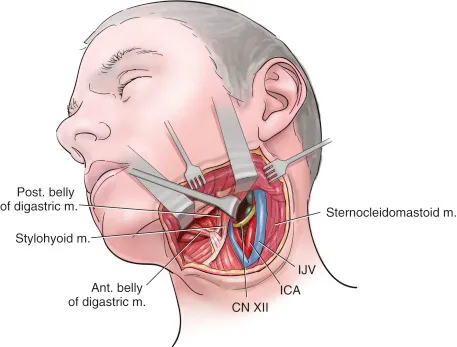
- Medial to internal carotid arteries: The paired retropharyngeal lymph nodes flank the medial sides of the internal carotid arteries bilaterally. The internal carotids are major arteries supplying blood to the brain.
- Medial and lateral masses: Each paired lymph node is organized into smaller medial and lateral masses.
- Lateral nodes medial to carotid sheath: The lateral mass of retropharyngeal lymph nodes lies just medial to the carotid sheath on each side. The carotid sheath envelops the internal carotid artery as well as internal jugular vein and vagus nerve.
- Posterior to buccopharyngeal fascia: This thin membrane separates the retropharyngeal lymph nodes from the pharynx anteriorly.
- Anterior to prevertebral muscles: The longus colli and longus capitis muscles lie posterior to the retropharyngeal space and nodes.
- Variable drainage: The retropharyngeal lymph nodes exhibit inconsistent drainage patterns. They often drain upward to the nodes below the skull base or downward to the jugulodigastric node at the junction of the internal jugular and digastric muscles.
- What Is the Survival Rate for Turner Syndrome?
- Does Elevating Your Legs Help with Circulation?
- Does Twinings Detox Tea Really Work?
Clinical Relevance of Retropharyngeal Lymph Node Location
Now that we have oriented the retropharyngeal lymph nodes anatomically, it is worth discussing their clinical significance:
- Lymphatic drainage: The retropharyngeal nodes drain various structures in the head and neck – from the nasal cavities, paranasal sinuses, soft palate, pharynx, larynx, thyroid gland, middle ear, and even the cerebrospinal fluid. This wide drainage explains their involvement in many pathologies.
- Metastatic cancer: Cancers of the head and neck commonly metastasize to the retropharyngeal lymph nodes. This includes malignancies of the nasopharynx, oropharynx, larynx, hypopharynx, and thyroid gland. Enlarged metastatic retropharyngeal lymph nodes can lead to symptoms like sore throat, dysphagia, and sleep apnea.
- Inflammation: The retropharyngeal nodes may become inflamed or infected due to regional infections like tonsillitis, otitis media, and retropharyngeal abscesses.
- Injury: Trauma to the cervical spine can also damage the retropharyngeal lymph nodes and lead to hemorrhage or edema.
Imaging to Identify Retropharyngeal Lymph Nodes
Radiologic imaging is key for visualizing retropharyngeal lymph nodes and assessing any abnormalities. Some modalities used include:
- MRI: Magnetic resonance imaging provides excellent soft tissue detail for visualizing lymph node anatomy and pathology.
- CT: Computed tomography allows clear views of the retropharyngeal space and lymph nodes when using contrast dye.
- Ultrasound: High-resolution neck ultrasound helps identify enlarged retropharyngeal lymph nodes. Doppler mode assesses lymph node vascularity.
- PET scan: Positron emission tomography combined with CT or MRI precisely locates metabolically active lymph nodes. This helps diagnose and stage cancers.
Correctly distinguishing enlarged metastatic lymph nodes from non-pathological asymmetries requires correlation with clinical findings. Images should be reviewed by specialized radiologists.
- Are Apples Good for You? Detailed Guide
- What Are the Most Survivable Cancers?
- How to Apply for Genesee Health Plan?
Surgical Approaches to Access the Retropharyngeal Lymph Nodes
The deep location of retropharyngeal lymph nodes necessitates meticulous surgical approaches:
- Transoral: Small metastatic nodes can be resected transorally, accessing the retropharyngeal space through an incision in the posterior pharyngeal wall. This avoids external scars.
- Transcervical: Suspicious enlarged nodes are often biopsied or excised via a horizontal neck incision at the appropriate cervical level. The carotid sheath is retracted laterally to access the retropharyngeal space.
- Transmandibular: Cancerous retropharyngeal nodes may require resection through a split mandible approach after mandibulotomy. This allows wide exposure.
- Endoscopic: Endoscopic transcervical approaches provide excellent visualization of the retropharyngeal space with minimal invasiveness. Robotic surgery also enables complex resections.
Precise knowledge of the relations and location allows safe access and avoids neurovascular injury during retropharyngeal lymph node procedures.
Retropharyngeal Lymph Node Location in Summary:
- Paired retropharyngeal nodes situated in buccopharyngeal fascia of retropharyngeal space
- Lie posterior to naso-oropharynx, anterior to C1 vertebra and longus capitis
- Found medial to internal carotids, divide into medial and lateral masses
- Lateral mass located medial to carotid sheath
- Drainage from head and neck structures makes them a site of metastases
- Imaging modalities like CT, MRI and ultrasound visualize nodes
- Surgical access to nodes achieved transorally, transcervically or with mandibulotomy
- Why Are Crystalloids Used in Sepsis?
- Which Immunoglobulin Is Present in Breast Milk?
- Should I Be Worried About Getting the COVID-19 Omicron Variant?
Conclusion:
Comprehensive knowledge of the anatomy and relations of the retropharyngeal lymph nodes is vitally important for physicians and surgeons treating head and neck pathologies. This article provided a detailed overview of the boundaries of the retropharyngeal space, fascial layers, adjacent neurovasculature, and typical drainage patterns that define the location of these lymph nodes. Additionally, it described relevant imaging modalities and surgical approaches used to access the retropharyngeal nodes. The intricacies covered will equip medical experts with a strong foundation for managing clinical issues related to the retropharyngeal lymph nodes confidently
- Why Exercise Is Important?
- Can You Take a Bath After a D&C?
- How Big Do Swordfish Grow?
- How to Clean Starpil Wax Warmer?
- How Many Brigades Are There in the 82nd Airborne Division?
- Why Are Dubia Roaches Illegal in Florida?
- Can BBQ Grills Go In A Dishwasher?
- Will Emily Wickersham Be Back on NCIS?
- How to Cut a Ping Pong Ball in Half?
- Why the King Needs a Secretary Chapter 49?
- Are Almonds Good for You?
- How Long Does Advil Gel Last? [Extend Life]
- Can You Wear Molded Cleats on Turf?
- Can You Sue Your Parents?
- How to Use Texblend to Fix Neck Seams in Skyrim?